Figure 3.
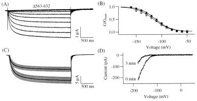
Properties of the Δ563–632 channel. (A) Representative current traces evoked in response to 2.5-s voltage pulses from −150 to −30 mV in 10-mV increments from a holding voltage of −10 mV. Tail currents were recorded at −60 mV. (B) Normalized G–V curves for KAT1 wild type (Δ, n = 5) and Δ563–632 channel (□, n = 6). (C) Comparison of the mean current amplitudes at −120 mV measured from the oocytes treated with colchicine-free ND96 solution (lower solid black line, n = 17) or with 100 μM colchicine-containing ND96 solution (upper solid black line, n = 18) for >22 h at 17°C. The shaded regions represent the corresponding SEM. (D) Current-voltage curves obtained by 2-s ramps from 50 to −190 mV were recorded in the inside-out configuration immediately (0 min) and 3 min after patch excision.

