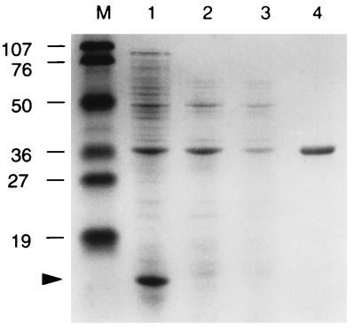
Figure 3.
SDS/PAGE analysis of bacterial surface proteins. An SDS/15% PAGE gel loaded with protein samples prepared from the surface of DC3000 (lane 1), hrcC mutant (lane 2), and hrpS mutant (lane 3) grown on solid hrp-inducing medium, or DC3000 grown on King’s medium B agar plates (lane 4). The gel was stained with 0.025% Coomassie brilliant blue R-250. Lane M, molecular mass markers (Bio-Rad) in kDa. Arrowhead indicates the 10-kDa protein.

