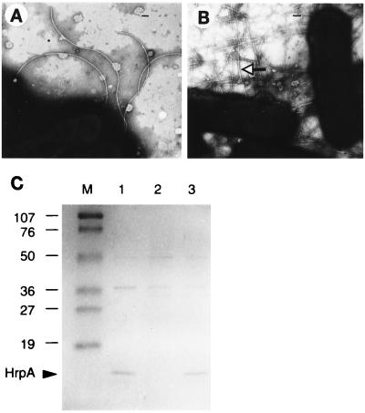
Figure 5.
Surface appendages and associated proteins on P. syringae pv. tomato DC3000 hrpA mutant and hrpA mutant containing pHRPA. (A) hrpA mutant and (B) hrpA mutant containing pHRPA grown on solid hrp-inducing medium were examined with a transmission electron microscope after staining with 1% potassium phosphotungstic acid (pH 6.5). Polar flagella of 15–18 nm in diameter are present on most cells of the hrpA mutant and the hrpA mutant containing pHRPA. Flagella were not seen in the field shown in B. In B, many Hrp pili of 6–8 nm in diameter are present (indicated by arrow). (Scale bars = 200 nm.) (C) An SDS/15% PAGE gel loaded with protein samples prepared from surface of DC3000 (lane 1), hrpA mutant (lane 2), and hrpA mutant containing pHRPA (lane 3) grown on solid hrp-inducing medium. Lane M, molecular weight markers (Bio-Rad) in kDa. The gel was stained with 0.025% Coomassie brilliant blue R-250.

