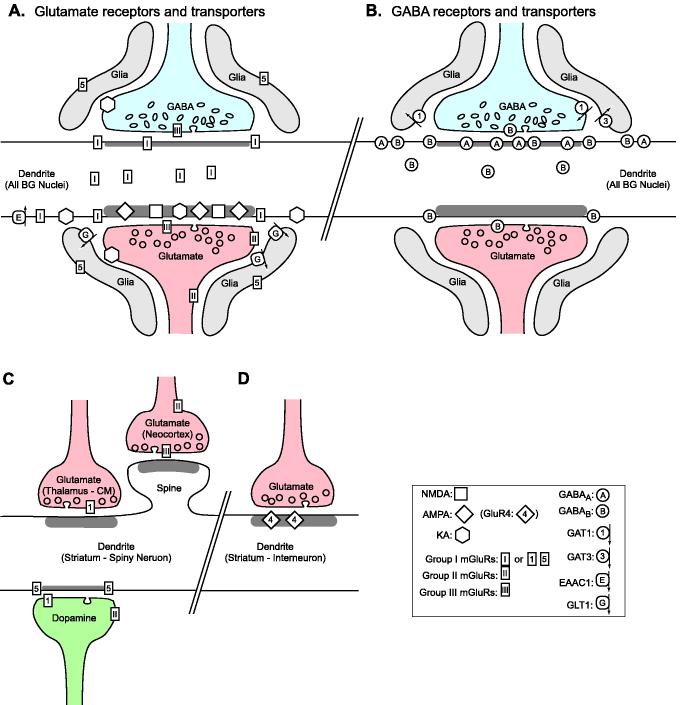
Fig. 7.
Summary figure of the subsynaptic localization of glutamate (panel A) and GABA (panel B) receptors and transporters in the basal ganglia, as revealed by immunogold EM studies. The schematic representation shows exemplary glutamate and GABA terminals that form axo-dendritic synapses. Most of the features depicted in A and B are valid for a majority of basal ganglia neurons; except for the spines of projection neurons and interneurons in the striatum, which are depicted in panels C and D respectively.

