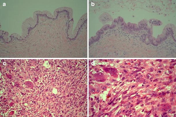
Fig. 2.

Cyst wall with typical tall columnar mucous-secreting epithelium and fibrous wall (a). Low cellular proliferation with some stratification of the cells and slight nuclear atypia (b). Heterogenous proliferation of pleomorphic cells, spindle cells, osteoclast-like giant cells, and some mononuclear cells with some pigment (c). Detail of sarcoma-like nodule (d)
