
Fig. 5.
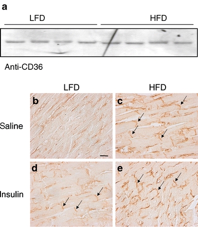
Expression and subcellular localisation of CD36. Expression of CD36 in ventricular lysates of HFD and LFD rats. Ponceau S staining confirmed that the filters contained equal amounts of ventricular extracts (a). Immunohistochemical staining for CD36 localisation of cardiac LV tissue sections from rats fed an LFD (b, d) or HFD (c, e) for 8 weeks. Rats received an i.p. injection of saline (b, c) or insulin (d, e) 30 min before killing. Photographs are representative of two independent experiments performed on three rats per experimental group. Arrows indicate intercalated discs. Scale bar indicates 25 μm