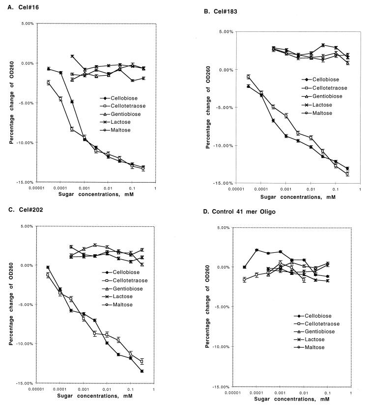
Figure 3.
Specificity of aptamer binding. Aptamers Cel#16, Cel#183, and Cel#202 were tested for the ability to bind four similar biose sugars by titrating the concentration of sugars and detecting DNA binding by measuring the resulting small decrease in absorbance of the solution at 260 nm (hypochromicity). The DNA concentration was constant within each experiment and was 0.6 to 0.7 μM. (A–C) For each aptamer, the cellobiose dimer and cellotetraose tetramer produced hypochromic shifts at similar concentrations (apparent Kd 10−7 to 10−5 M), but the related biose sugars did not produce significant hypochromicity. (D) A control 41-mer DNA oligonucleotide did not give a hypochromic response to any of the sugars tested.

