
Figure 4. Fur binds the iha promoter region.
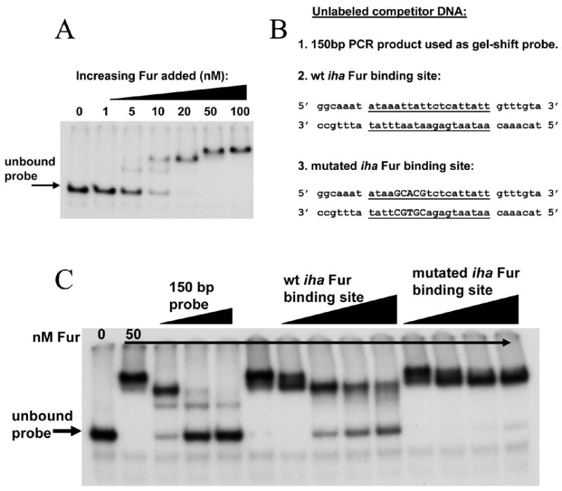
(A): A 150 bp DNA fragment encompassing the putative Fur-binding site was radiolabelled, incubated with increasing concentrations of Fur protein, and separated on a 5% acrylamide gel.
(B): Unlabeled competitor DNA sequences. Center 19 nucleotides represent the putative iha Fur binding site (underlined). Capital letters represent sequence that has been changed to decrease Fur binding.
(C): Fur binding in the presence of unlabeled competitor DNA. Lane 1, probe only; all other lanes, probe with 50 nM Fur. Lanes 3–5 contain 0.5 pMol, 2 pMol, and 5 pMol unlabeled probe, respectively. Lanes 7–10 contain 10 ng, 50 ng, 100 ng, and 150 ng, respectively, of unlabeled wt iha Fur binding site. Lanes 11–14 contain 10 ng, 50 ng, 100 ng, and 150 ng, respectively, of unlabeled mutated iha Fur binding site.