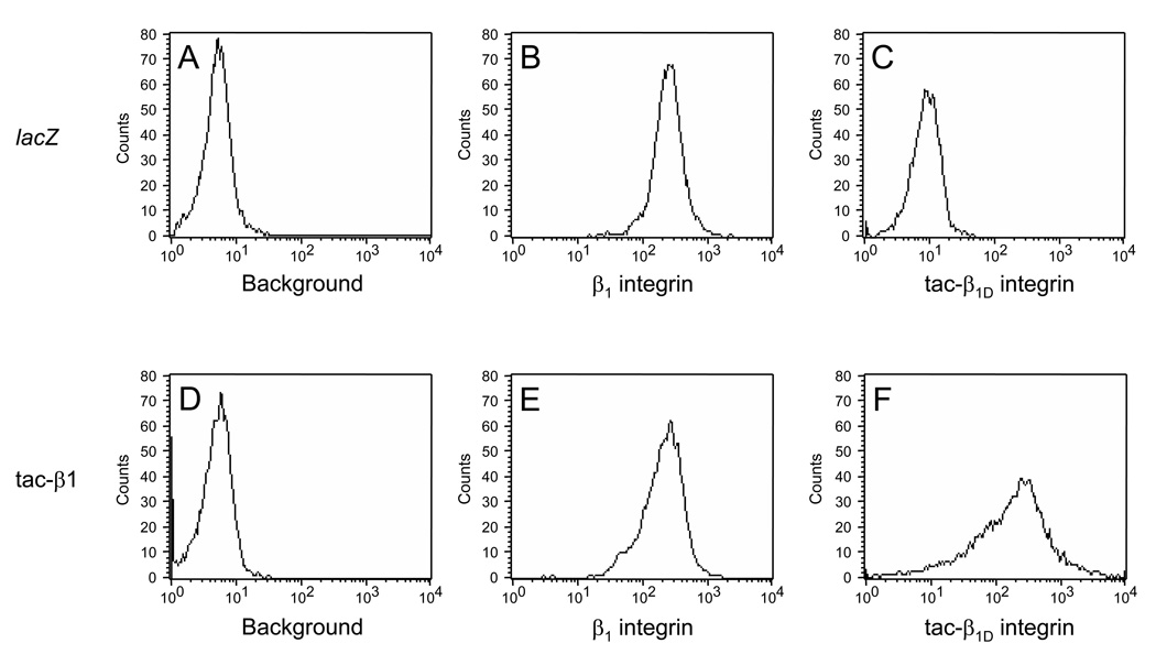
Fig. 2. Expression of β1 integrin and tac-β1 in transfected cells.
The presence of cell surface β1 integrin was verified by staining cells with FITC-conjugated β1 integrin antibody 24 h after infecting cells with adenovirus expressing lacZ (B, MFI = 264 ± 0.55) or tac-β1 (E, MFI = 240 ± 1.94). To determine expression levels tac-β1 cells were initially incubated with IL-2α receptor antibody, which detects an epitope (residues 140–144 of human IL-2 receptor α) in the extracellular domain of tac-β1 protein. Surface expression of tac-β1D was measured by detecting secondary antibody conjugated to Alexa-488, which was absent in cells infected with adenovirus expressing lacZ (C, MFI = 10.6 ± 0.07), compared to tac-β1D chimera (F, MFI = 326 ± 6.05). Background fluorescence in NRVM expressing lacZ (A, MFI = 5.17 ± 0.03) and tac-β1D (D, MFI = 5.35 ± 0.03) were determined by incubating cells in the absence of antibody. The mean fluorescent intensity and standard error (MFI ± SEM) for each distribution were obtained from a sample of 10,000 cells.

