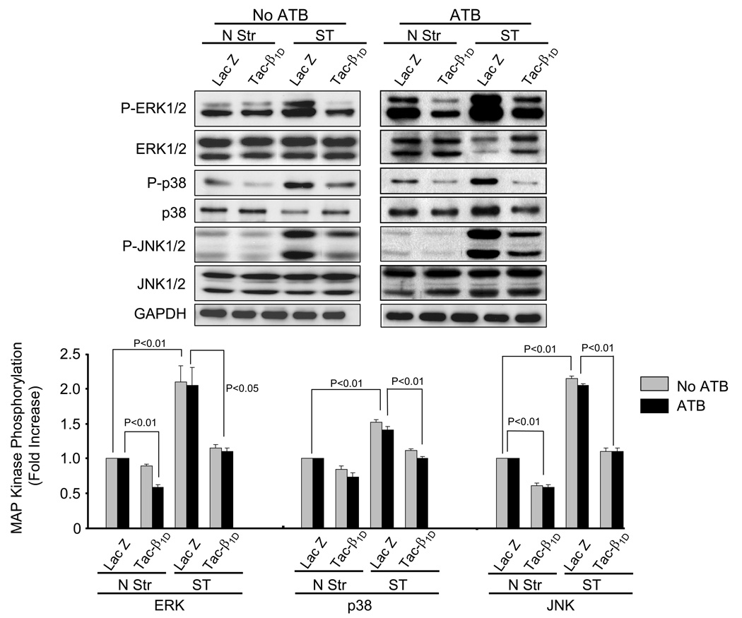
Fig. 3. β1D integrin is required for stretch-induced phosphorylation of ERK1/2, p38, and JNK.
Prior to stretching, NRVM were infected with adenovirus (24 h) which resulted in expression of tac-β1D or control protein (lacZ). After 5 min of stretch (Str) or no stretch (N Str), phosphorylation levels of ERK, p38 and JNK were determined in the presence and absence of ATB. Bar graphs show fold changes in MAP kinase phosphorylation after tac-β1D expression compared to control virus. Values are means ± SEM. N=5 experiments.

