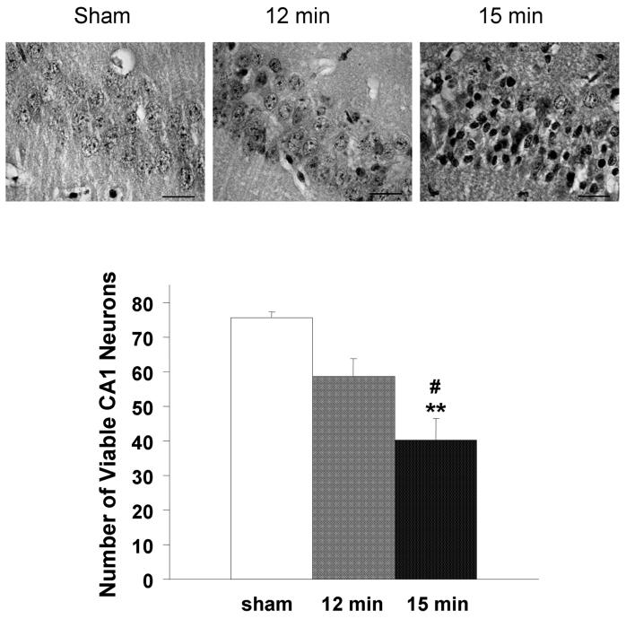
Fig. 1.
Neuronal loss after different ischemic durations. The number of viable CA1 neuronal cells was counted under a light microscope. Pyknotic eosinophilic neurons indicated ischemic damage. Neuronal damage was expressed as the mean number of surviving neurons in the CA1 subfield of each observed field (100 × oil lens). The number of viable neurons in hemispheres exposed to 15-min ischemia followed by 7-day reperfusion (n = 40) was significantly less than that of hemispheres from sham-operated mice (n = 16) and mice that underwent 12-min ischemia (n = 16). There were no significant differences between the 12-min ischemia group and the sham-operated group. **p<0.01 vs sham, #p<0.05 vs 12-min.

