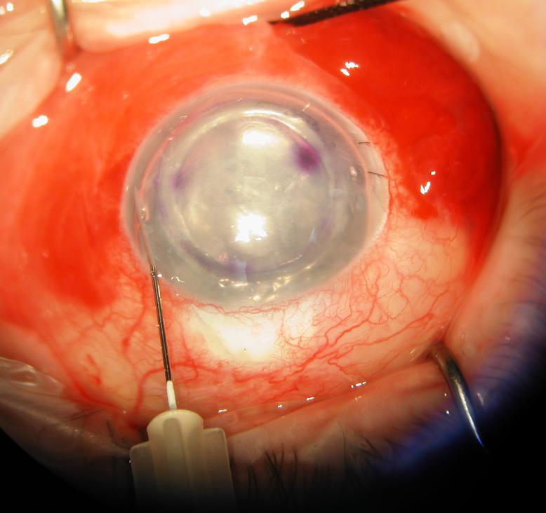
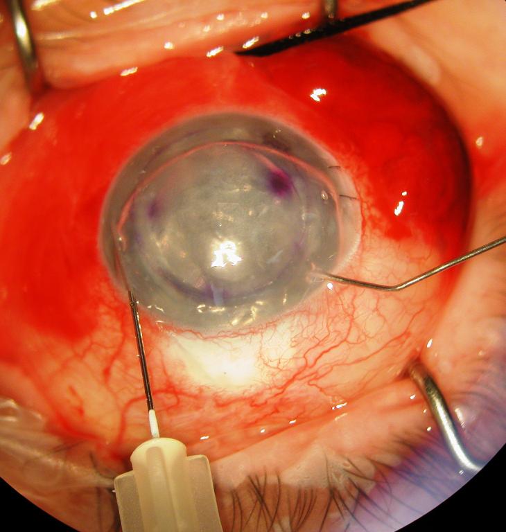
Figure 1.
Surgeon's view of the right eye depicting an infusion/air–fluid exchange technique for controlling graft tamponade pressure during DSAEK. The posterior lamellar graft is shown in its final position with a subtotal air fill maintained at 30 mm Hg by the Accurus surgical system. The system is connected via tubing to a 30-gauge needle that is inserted through a long nasal corneal tract (left). After 10 to 15 minutes, a partial air–fluid exchange is begun while the tamponade pressure is maintained (right). A manual infusion cannula is used to separate the wound margins of a paracentesis to allow controlled escape of air during the exchange.


