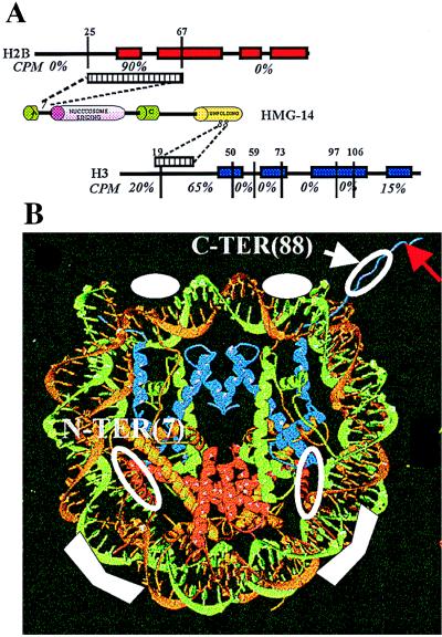
Figure 6.
Organization of HMG-14 in nucleosome cores. (A) Specific crosslinks between HMG-14 and histones in nucleosome cores. The α-helical regions in the histones are depicted as boxes. The evolutionarily conserved domains in HMG-14 are depicted as cylinders. The amino acid positions at which the proteases cleaved the histones are indicated above their sequence. The radioactive counts present in each of the peptides is indicated, expressed as percent of total. The regions targeted in H2B and H3 by amino acid residues 7 and 88 of HMG-14, respectively, are indicated by the striped boxes. (B) Model of the HMG-14 binding sites. The ribbon traces for the DNA and histones in the core particles were reproduced by permission from the recent article in Nature by Luger et al. (12), copyright 1997, Macmillan Magazines Ltd. The solid white symbols, in the two major grooves flanking the dyad axis and approximately 25 bp from the end of the DNA, indicate the regions where HMG-14/-17 protect the DNA from hydroxyl radical cleavage. The open white circles represent the approximate location of the crosslinks identified in the present study. The red arrow points to the N-terminal region of histone H3. Histones H2B and H3 are represented by red and blue ribbons, respectively.

