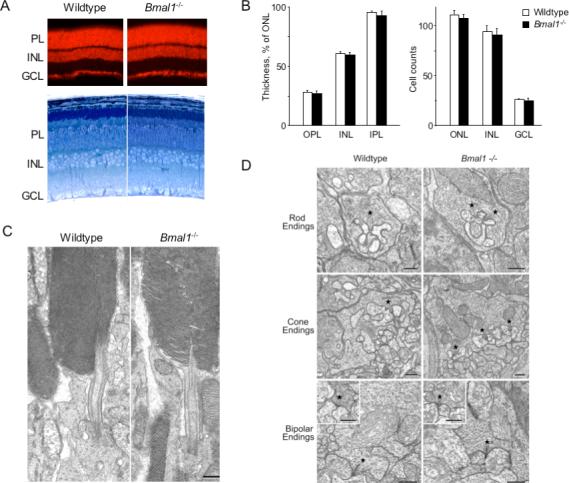
Figure 2. Normal retinal architecture, cellular organization, and ultrastructure in Bmal1−/− mutant mice.

(A) Top, fluorescence images of retina sections from adult littermate wildtype or Bmal1−/− mutant mice stained with ethidium bromide to show cell nuclei. Organization of nuclear layers was indistinguishable in the two genotypes. Bottom, thick plastic sections of retinas from adult littermate wildtype or Bmal1−/− mice stained with toluidine blue to show cell morphology and structure. No difference between genotypes was observed. PL, photoreceptor layer; INL, inner nuclear layer; GCL, ganglion cell layer. (B) Quantitative comparison of thickness (left) and cell densities (right) (mean and SEM; N = 3) of retinal layers (at mid-periphery) of adult wildtype and Bmal1−/− littermate mice (Experimental Procedures). Results for central retina were similar (data not shown). There were no significant differences between genotypes. OPL, outer plexiform layer; IPL, inner plexiform layer. Cell counts: for ONL, INL--per 5000-μm2 area; GCL--per 200-μm segment of retina. (C) Representative electron micrographs of rod photoreceptors from adult littermate wildtype or Bmal1−/− mice. No difference between genotypes was observed in the fine structure of outer and inner segments. (D) Representative electron micrographs from adult littermate wildtype or Bmal1−/− mice. No differences between genotypes were evident in the morphology of synaptic endings of rods, cones, rod bipolars, or cone bipolars (insets) or in the complement of synaptic vesicles or structure of ribbon synapses (asterisks). (C,D) Scale bars, 500 nm.
