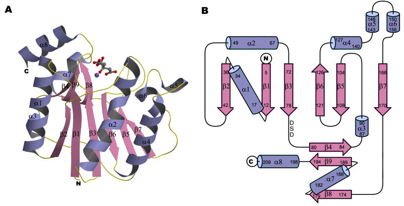
Figure 2. Crystal structure of the glycosyltransferase domain of A64R.
(A) Ribbon diagram with bound Mn2+ and citrate ions. Mn2+ and citrate ions are represented in ball-and-stick representation. N, C, O, and Mn atoms are colored blue, grey, red, and purple, respectively.
(B) Topology diagram of the A64R structure. The DXD motif is indicated.

