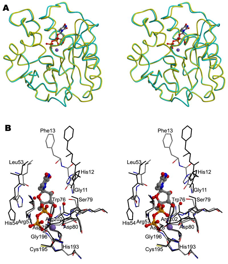
Figure 3. The structure of A64R complexed with UDP.

(A) Stereodiagram showing the superposition of the Cα traces of A64R with (yellow) or without UDP (cyan) bound. UDP is shown in a ball-and-stick representation and colored the same as in Figure 2. P atoms are colored in orange.
(B) Stereodiagram showing the interaction between UDP/Mn2+ and protein residues within 4 Å of UDP. Water molecules which form hydrogen bonds with UDP are also shown. UDP is shown in a ball-and-stick representation. The structure of A64R when UDP bound is color-coded according to the nature of the atoms. The A64R structure without bound UDP is colored black.
