
Figure 1.
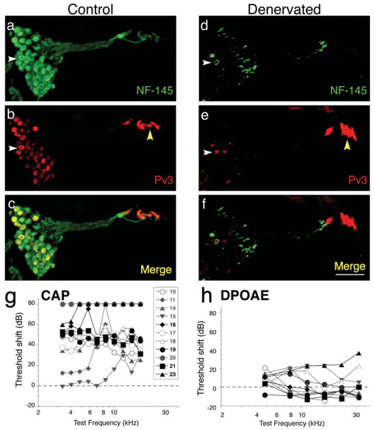
Denervation of the gerbil cochlea. Cochlear sections from a control ear (a–c) and a ouabain-treated ear (d–f; referred to as denervated in all figures) show primary neuronal degeneration 8 days after treatment. Sections were stained by immunohistochemistry for neurofilament (green) and parvalbumin 3 (red). Neurofilament-positive spiral ganglion neurons are plentiful in Rosenthal’s canal in the control ear (white arrowhead in a) but are almost completely absent in the denervated ear (white arrowhead in d). Parvalbumin 3 also stains most neuronal cell bodies (white arrowhead in b), as well as the hair cells (yellow arrowhead in b). In the section from the denervated ear, hair cells remain intact (yellow arrowhead in e) but the spiral ganglion neurons are almost completely absent (white arrowhead in e). Merged images are shown (c and f) and the scale bar in f is 80 μm and applies to a–f. (g and h): Compound action potential (CAP) thresholds (g) were significantly elevated, consistent with massive loss of spiral ganglion cells, whereas distortion product otoacoustic emissions (DPOAEs) were close to normal (h), indicating that cochlear hair cells remained healthy. Data from eleven cases are shown; the symbol key in g also applies to h. Highlighted black symbols in g and h are the cases that received long-term grafts. Threshold shift for each measure was computed by subtracting the data from each case from the mean values for all ears obtained before the ouabain treatment. [Color figure can be viewed in the online issue, which is available at www.interscience.wiley.com.]