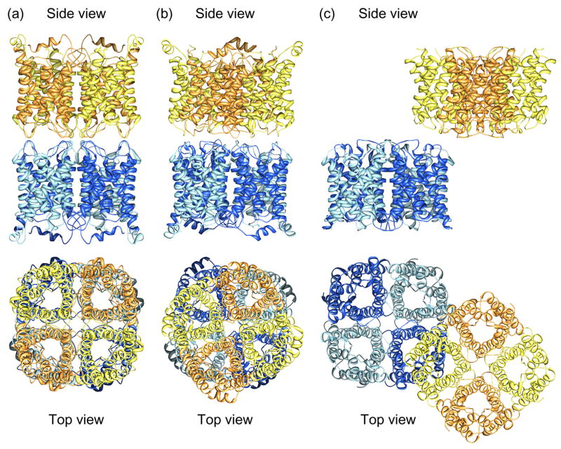
Figure 1.
Membrane junctions formed by aquaporins-0 and 4. (a) Structure of the aquaporin-0 mediated membrane junction visualized by electron crystallography of double-layered 2D crystals (pdb accession code: 2B6O) [10,11●●]. The paired tetramers are exactly in register. (b) Paired aquaporin-0 tetramers as seen in loosely packed 3D crystals (pdb accession code: 2C32) [37]. The paired tetramers are rotated by 24° with respect to each other. (c) Structure of the aquaporin-4 mediated membrane junction visualized by electron crystallography of double-layered 2D crystals (pdb accession code: 2D57) [12●●].

