Figure 1.

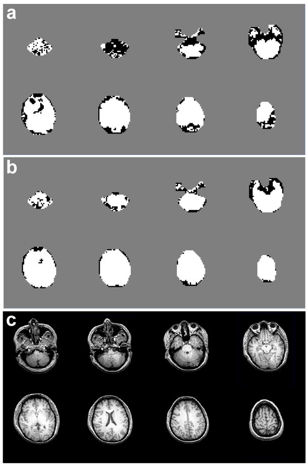
Maps of accepted voxels (white) and rejected voxels (black) in one volunteer studied at 4 T (TE=45 ms) without (a) and with local B0-shift correction (b). Also shown are the corresponding structural MR images (c).

Maps of accepted voxels (white) and rejected voxels (black) in one volunteer studied at 4 T (TE=45 ms) without (a) and with local B0-shift correction (b). Also shown are the corresponding structural MR images (c).