
Figure 3.
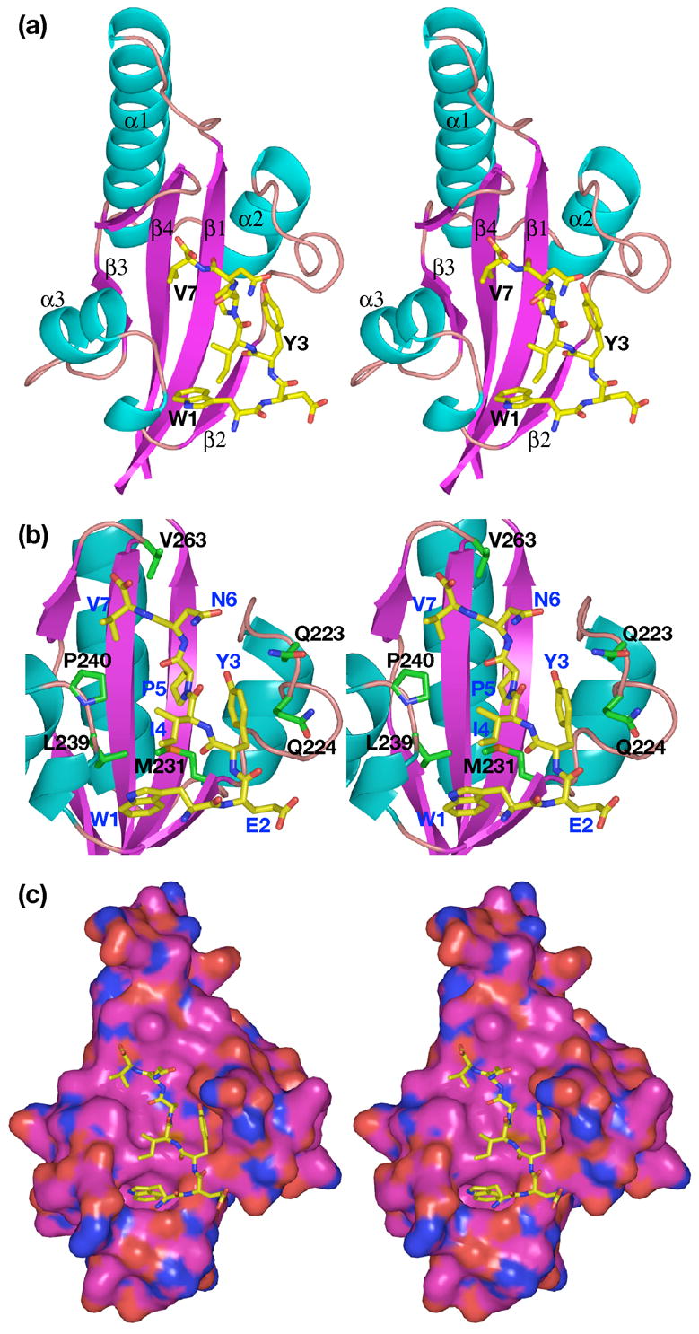
Stereo views of the complex of the N-peptide with SurA-P1. (a) Ribbon drawing of the complex. α-helices and strands of β-sheet are numbered sequentially; note that Pro258 introduces a break in β3. Helices are cyan; β-strands, magenta; oxygen atoms, red; nitrogen atoms, blue; carbon atoms, green on protein, yellow on peptide. (b) View showing interactions between residues on peptide and protein. Protein residues are labeled in black; peptide residues labeled in blue. Colors are same as in (a); additionally, sulfur atoms are orange. (c) Space-filling surface of protein. Colors as above, except carbon atoms of protein are magenta.