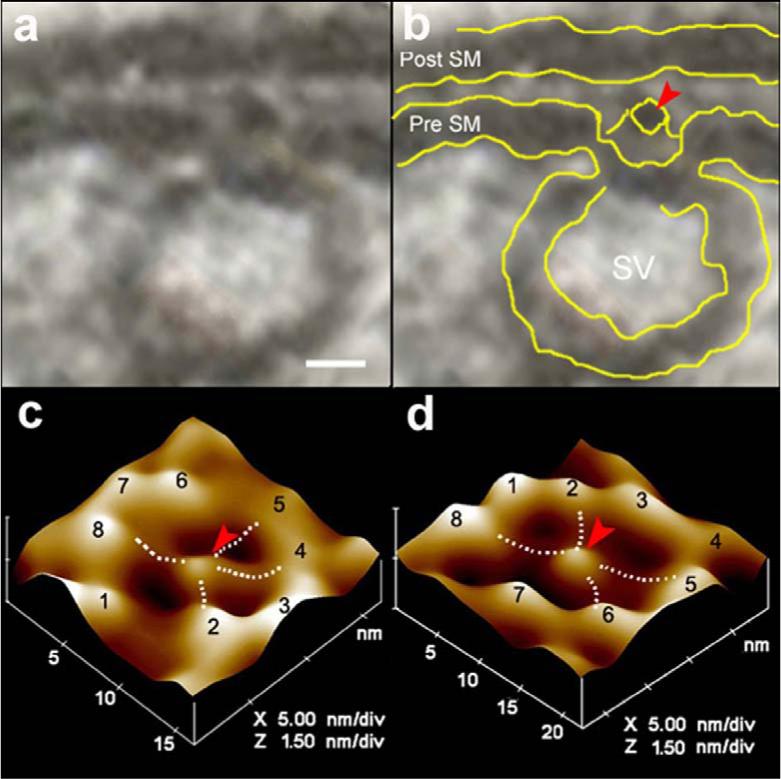
Figure 1. Neuronal fusion pore.

(a,b) Electron micrograph of a cup-shaped neuronal fusion pore measuring 12 nm at the presynaptic membrane, where a 30 nm synaptic vesicles is docked. Note the central plug (red arrowhead) of the fusion pore complex. Bar = 5 nm. (c) Atomic force micrograph of the surface topology of a neuronal fusion pore at the presynaptic membrane in an isolated synaptosome. (d) Atomic force micrograph of an isolated neuronal fusion pore, reconstituted in lipid membrane. Note the central plug and the 8 ridges in the atomic force micrographs of the fusion pore, both at the presynaptic membrane of the synaptosome (c) and in the isolated lipid-reconstituted complex (d). Bridges connecting the ridges with the central plug (dotted lines) are seen in the atomic force micrographs.
