
Figure 3. LCQ quantitation of tandem MS spectra of in vitro unlabeled and labeled Aβ17-28.
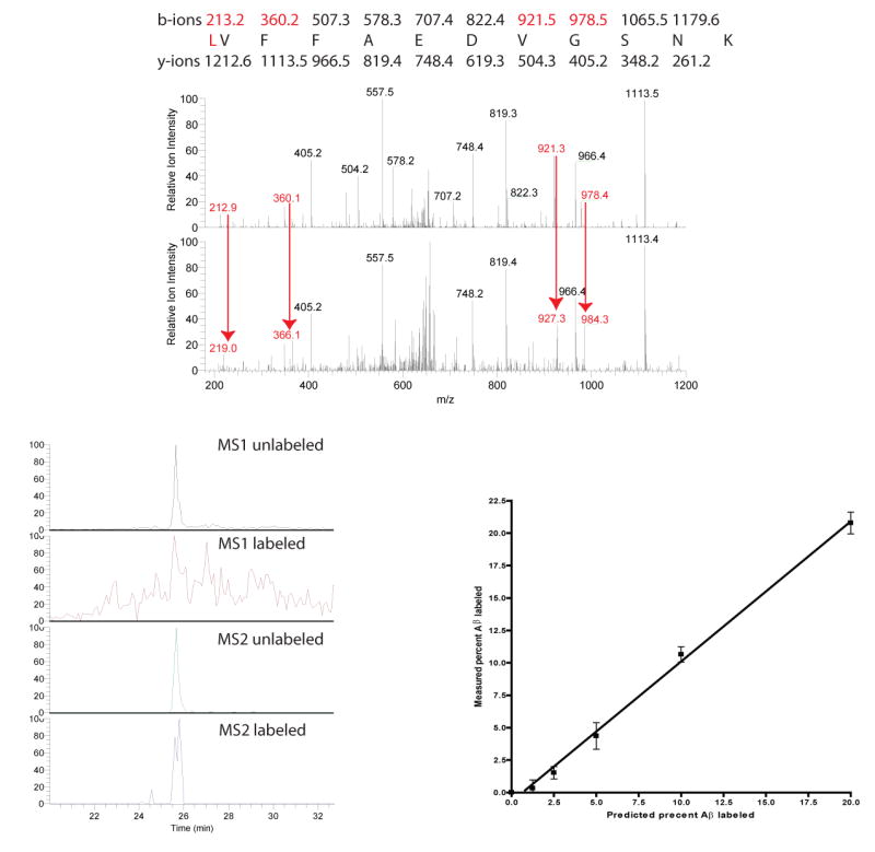
(a) Neuroglioma cell media that was unlabeled (top) or labeled (bottom) with 13C6-leucine. The spectra were obtained using tandem MS analysis of unlabeled parent ion Aβ17-28 (m=1325) or labeled parent ion Aβ17-28(m=1331) by LCQ-ESI-MS. Note the tandem MS ions containing leucine at Aβ17 (see masses 213, 360, 921, and 978) are mass shifted by 6 Daltons demonstrating the labeled leucine (arrows). The Aß ions without leucine are not labeled and are not mass shifted by 6 Daltons (see mass 348.2 and 405.3 in both spectra).
(b) Base peak chromatograms of MS1 versus MS2 quantitation. 2.5% leucine labeled culture was analyzed in the same MS run after immunoprecipitation and trypsin digestion of Aβ. Reconstructed base peak chromatograms demonstrate the signal to noise ratio improvements of MS2 versus MS1 quantitation.
(c) Standard curve of labeled Aß to unlabeled Aß. Labeled cultured media was serially diluted with unlabeled media to generate samples for a standard curve. Aß was immuno-precipitated from the media, trypsin digested, and Aβ17-28 fragments were analyzed on a LCQ-ESI-MS and the tandem mass spectra ions were quantitated using custom written software. The predicted percent labeled Aß versus the measured value is shown with a linear regression line.