
Fig. 2.
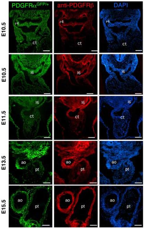
PDGFRα and PDGFRβ expression in the cardiac outflow tract PDGFRα expression was tracked using embryos that possessed a PDGFRαGFP allele and PDGFRβ was examined using immunohistochemistry. All three images at each stage are the same section. Embryonic stage is indicated at the left. Two E10.5 sections were imaged to illustrate PDGF receptor expression during migration of the cardiac NCC. r4, right fourth arch artery; l6 left sixth arch artery; ct, conotruncus; ao, aorta; and pt, pulmonary trunk. Note that the PDGFRα expression is followed using a nuclear localized GFP. Scale bar: 200μm.