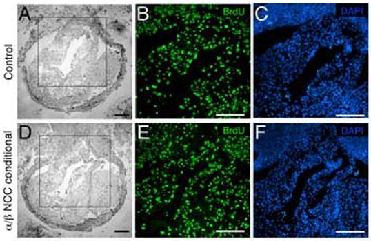
Fig. 6.

NCC proliferation remains unchanged in the outflow tract of PDGFRα/β NCC conditional mutants Transverse sections of E10.5 aortic sac stained for BrdU incorporation. (A-C) Control (PDGFRαfl/+; PDGFRβfl/+; Wnt1-CreTg) and (D-F) PDGFRα/β NCC conditional (PDGFRαfl/fl; PDGFRβfl/fl>;Wnt1-CreTg) embryos. (A, D) Brightfield images. Boxed region indicates close-up area for fluorescence detection. (B, E) BrdU positive nuclei and (C, F) DAPI stained nuclei. (ao) aorta, (es) esophagus (tr) trachea, (ta) truncus arteriosus. Scale bar: 200μm.
