Fig. 7.

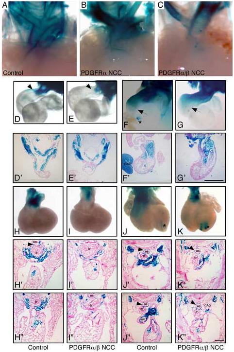
Reduced NCC migration into the conotruncus Detection of NCCs via the R26R-LacZ/Wnt1-CreTg lineage marker. Whole mount β-galactosidase activity of aortic arches at (A-C) E16.5, (D, E) E10.5, (F, G) E11.5, (H, I) E12.5, (J, K) E13.5. Asterisk indicates aberrant Wnt1-Cre activity that has been previously reported (Stottmann et al., 2004). Arrows point to furthest migration point of β-galacotosidase-tagged NCC. (D’-K’ and H”-K”) figures were transverse sections from control and mutant embryos. E10.5 and E11.5 sections were generated from the whole mount stained embryos. E12.5 and E13.5 were independent embryos that were frozen embedded, sectioned, and stained for β-galactosidase activity at the level of the outflow tract. Single prime (’) images were anterior to the double prime (”) images. A marked reduction in NCCs in the aortico-pulmonary septum of the aortic arch can be observed in the PDGFRα/β NCC conditional embryos at E12.5. Genotypes are as indicated. For whole mount images surrounding tissues were removed from the embryos to enhance visualization of the aortic arch region. BA2; second branchial arch; es, esophagus; Ao, aorta; dAo, descending aorta; and tr, trachea.
