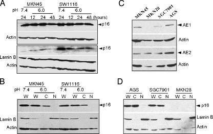
Figure 3.
The cytoplasmic distribution of p16 in MKN45 and SW1116 cells was not affected by the low pH of the culture, and expression occurred in the three other gastric adenocarcinoma cell lines. (A) p16 expression in whole-cell extract (upper panel) and nucleus (lower panel) from two tumor cell lines cultured at pH 6.0 (for 12, 24, and 48 hours) and pH 7.4. (B) Direct comparison of the nuclear-cytoplasmic distribution of p16 in the two tumor cell lines cultured at pH 6.0 and 7.4. (C) The expression of AE1 and AE2 in the three other gastric adenocarcinoma cell lines. (D) Cytoplasmic distribution of p16 in the three other gastric adenocarcinoma cell lines. W, whole-cell extract; C, cytoplasmic fraction; N, nuclear fraction. MKN28, AGS, and SGC7901 represent well, moderately, and poorly differentiated gastric adenocarcinoma cell lines, respectively.

