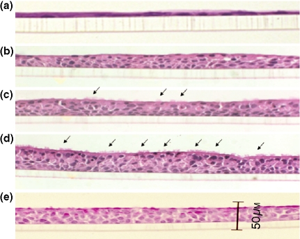
Figure 1.
Histological appearance of mucociliary differentiation of NHNE cells over time. At the time of confluence, there was only a monolayer of cells (a). On the 7th day after confluence, the cells grew to form several layers (b). On the 14th day after confluence, ciliated cells could occasionally be seen (c). On the 28th day after confluence, the amount of ciliated cells was greater and the cells themselves became more cuboidal (d). To see whether these cells were mucous cells, they were stained with PAS solution. Many cells containing mucus could be observed (e). Arrows indicate the ciliated cells.

