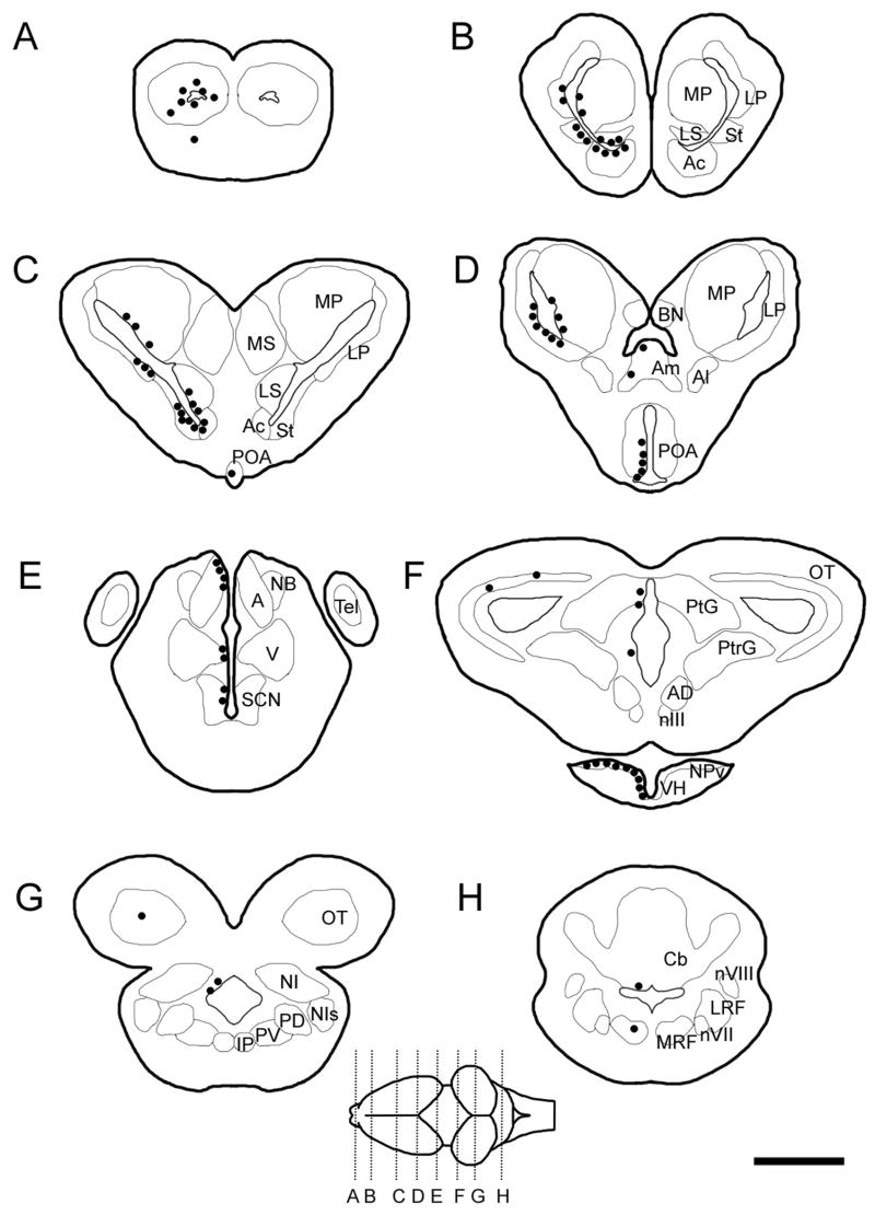
Figure 1.
Schematics of brain sections showing the distribution of BrdU+ cells (black circles) at 2–48 hours. (ABBREVIATIONS: A, anterior thalamic nucleus; Ac, nucleus accumbens; AD, anterodorsal tegmentum; Al, lateral amygdala; Am, medial amygdala; BN, bed nucleus of the pallial commissure; Cb, cerebellum; Ip, interpeduncular nucleus; LP, lateral pallium; LRF, lateral reticular formation; LS, lateral septal nucleus; MP, medial pallium; MRF, medial reticular formation; MS, medial septal nucleus; NB, nucleus of Bellonci; NI, nucleus isthmi; NIs, secondary isthmal nucleu; NPv, nucleus of the periventicular organ; nIII, oculomotor nucleus, nVII, vestibular nucleus; nVIII, facial motor nucleus; OT, optic tectum; PD, posterodorsal tegmentum; PV, posteroventral tegmentum; PO, preoptic area; PtG, pretectal gray; PtrG, pretoral gray; SCN, suprachiasmatic nucleus; St, striatum; V, ventral thalamus.)

