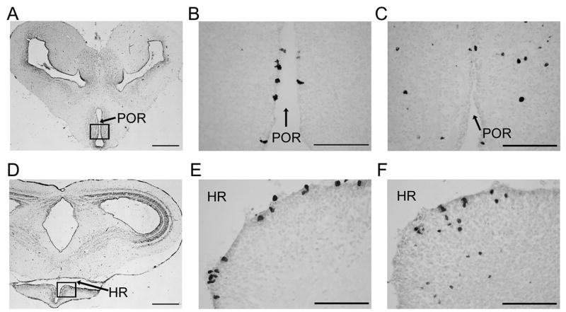
Figure 2.
Photomicrographs of the anuran amphibian telencephalon: Nissl-stained sections (A, D) with squared regions containing BrdU-labeled cells (B, C, E, F). Preoptic area depicted in top row and ventral hypothalamus in bottom row (preoptic recess, POR; hypothalamic recess, HR). BrdU+ cells are in the ependymal layer at 48 hrs (B, E) and in the parenchyma at 30 days (C, F). (Scale bar in A, D = 500 μm; scale bar in B, C, E, F = 100 μm).

