Figure 1.

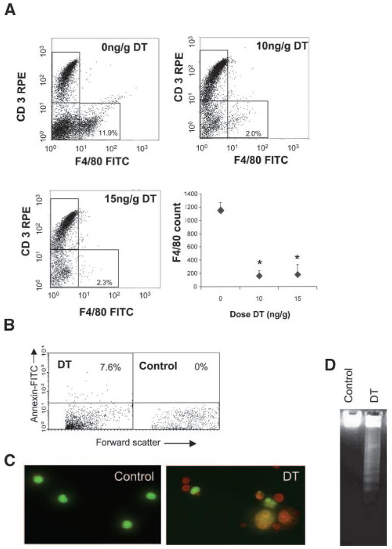
Characterization of CD11b-DTR mice. A, Representative flow-cytometric profiles from DTR-FV/B mice treated with 2 doses of 10 ng/g or 15 ng/g DT at 48-hour intervals. Blood was isolated 24 hours later, labeled with F4/80–fluorescein isothiocyanate (FITC) and CD3-R-phycoerythrin (CD3-RPE) antibodies and analyzed by flow cytometry. Absolute F4/80 counts are also shown for each dose of DT as mean±SEM. *P<0.05 (n=3) compared with controls (0 ng/g DT). B, Flow cytometry for Annexin V demonstrating increased circulating Annexin V–positive cells after DT treatment. C and D, Peritoneal macrophages isolated from these mice demonstrate apoptosis on acridine orange staining (orange cells with condensed chromatin) (C) and DNA laddering (D).
