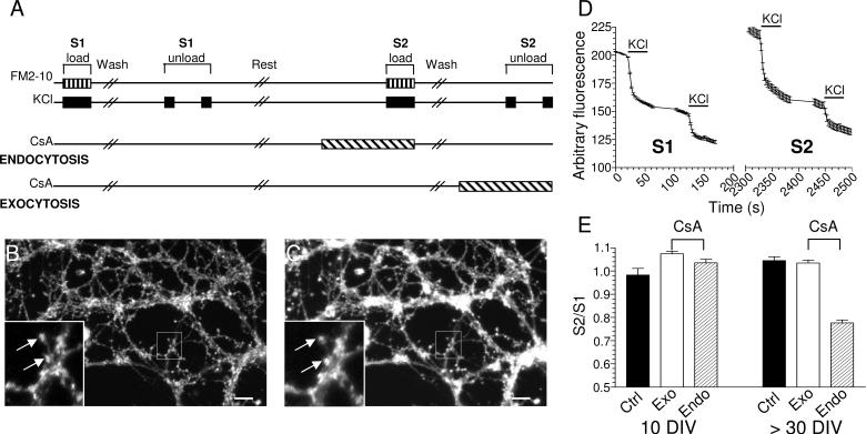
Fig 5. SV endocytosis develops a CaN requirement with age in culture.
(A) Protocol for quantifying the effect of CsA on SV exocytosis and endocytosis in CGNs. Image of CGN neurite field from a 10 days in vitro (DIV) culture loaded with FM2-10 at S1 (B) and S2 (C). Inset displays the reproducible loading of nerve terminals during repetitive stimuli (arrows). Scale bar denotes 1 μm. (D) Example of FM2-10 unloading from 10 DIV nerve terminals loaded at S1 and S2. The fluorescence decrease from 2 repetitive 50 mM KCl stimuli (bars) are summed and taken as the total amount of accumulated FM2-10 at S1 or S2 respectively. (E) S2 / S1 ratio of the effect of CsA (40 μM for 15 min) on either SV exocytosis (open bars) or endocytosis (hatched bars) in young (10 DIV) or mature (> 30 DIV) cultures. Experiments were ≥3 (10 DIV - control n = 221, CsA exocytosis n = 345, CsA endocytosis n = 246; > 30 DIV - control n = 252, CsA exocytosis n = 351, CsA endocytosis n = 215; all ± SEM).

