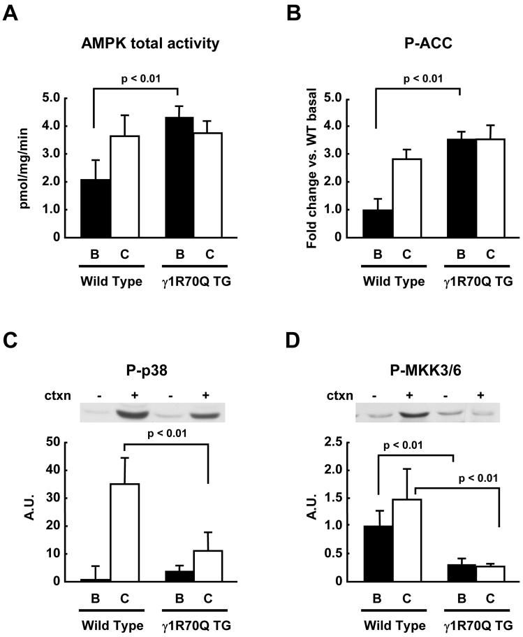
Figure 3. p38 MAPK phosphorylation in γ1R70Q TG mice.
Wild type and γ1R70Q TG mice were anesthetized, and the sciatic nerves were attached to electrodes. One leg was electrically stimulated for 10 min to induce muscle contractions, and the other leg served as sham control. Gastrocnemius muscles were dissected. In vitro kinase assay was done to determine total AMPK activity after immunoprecipitation with an antibody that recognizes both α1 and α2 AMPK isoforms (A). The same protein samples used for the kinase assays were used for immunoblotting of phospho-ACC (B), phospho-p38 MAPK (C), and phospho-MKK3/6 (D). Data are the means ± SEM. n = 6/group.

