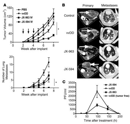
Figure 8. Rabbits bearing VX2 tumors implanted into the liver were followed by CT imaging at the indicated times after tumor implantation.
(A) 1 × 10⁸ PFU of viruses JX-594 (Wyeth strain, TK-deleted, expressing human GM-CSF), vvDD, or JX-963 (vvDD–GM-CSF) were delivered by ear vein injection at 2, 3, and 4 weeks after tumor implantation (arrows), when tumors measured approximately 5 cm3. The primary tumor burden (top) and the number of detectable lung metastases (by CT scanning; bottom) were measured (n = 18 for control-treated animals; n = 6 for vvDD-treated; n = 6 for JX-963–treated) *P < 0.05. (B) Representative CT scans of the primary tumor in the liver (left panels; tumor indicated by arrows) and metastases in the lungs (right panels) at 6 weeks after implantation. (C) Animals were also bled at the indicated times after treatment, and the numbers of viral infectious units (PFU/ml) were titered in the blood .

