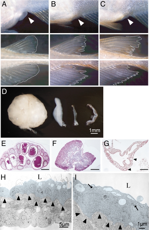
Fig. 2.
Sex reversal of germ-cell-deficient medaka and its associated gonadal morphology. (A--C) Representative images of the secondary sex characteristics of medaka adults that are genetically female (XX) (A), male (XY) (B), and cxcr4-morphant (germ-cell-deficient) female (XX) (C). (Top) External genitalia. White arrowheads indicate the anus. A developed urinogenital papilla is observed only in the wild-type female. (Middle and Bottom) Dorsal and anal fins, respectively. Phenotypic males (B and C) display a sharp and long anal fin, whereas a round-shaped anal fin is characteristic of a phenotypic female (A). (D) Dorsal views of an ovary (left), testis (second from left) and germ cell-deficient XX and XY gonads (second from right and right). (E–G), Cross-sections of ovary (E; scale bar, 500 μm), testis (F; scale bar, 200 μm), and germ-cell-deficient gonad (G; scale bar, 100 μm). A single empty lumen with several foldings is present in the germ-cell-deficient gonad. Arrowheads indicate blood vessels. (H and I) Electron micrographs of germ-cell-deficient medaka gonads. The lumen (L), basement membrane (arrowheads), and desmosomes (arrows) are indicated. A single layer of innermost cells is shaded with a pale blue color.

