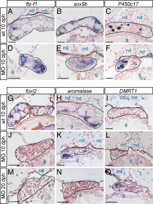
Fig. 4.
Gene expression analysis of both wild-type and germ-cell-deficient developing gonads. The XX gonads of 10 dph wild-type (A–C and G–I), 10 dph morphant (D–F and J–L), and 20 dph morphant (M–O) were analyzed by in situ hybridization for the marker genes, which are indicated on the top of columns. Nuclei are counterstained with neutral red. The gonad in each image is enclosed by a black dotted line. Nephric duct (nd), gut (g). (Scale bars, 20 μm.)

