Fig. 5.
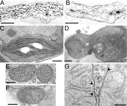
Leaf morphology and ultrastructure of mgd1-2 plastids. (A and B) Light microscopy of WT (A) and mgd1-2 mutant (B) leaf sections. (Scale bars: 100 μm.) (C and D) Electron micrographs of plastids from WT (C) and mgd1-2 (D) leaves. (Scale bars: 1.0 μm.) (E and F) Electron micrographs of mitochondria from WT (E) and mgd1-2 (F). (Scale bars: 0.2 μm.) (G) A close-up of the membrane structures indicated by an arrow in D. Arrowheads indicate the sites of inner envelope invagination. (Scale bar: 0.2 μm.)

