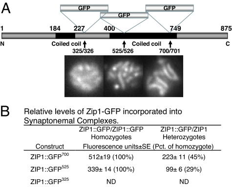
Fig. 1.
Structure of ZIP1::GFP constructs and relative efficiency of their incorporation into SCs. (A) ZIP1 gene showing location of GFP inserts. Constructs were introduced into S. cerevisiae as described in Materials and Methods. Representative fluorescent images of pachytene nuclei for each construct are shown below. (B) Relative efficiency of incorporation into SCs in heterozygotes and homozyotes. Strains HW122, HW123, EW102, and EW103 were incubated in sporulation medium and analyzed when the percentage of cells in pachytene was highest. Zip1 fluorescence was quantitated as described in Materials and Methods. ND, not determined.

