Fig. 3.
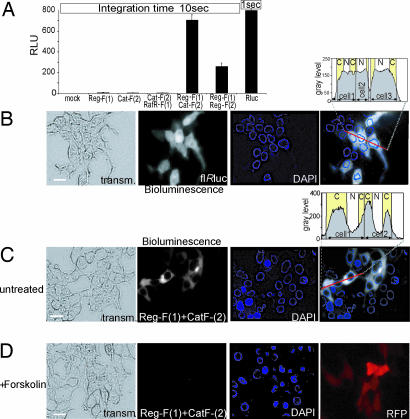
Cellular imaging of bioluminescence of transiently transfected HEK293T cells expressing full-length Rluc and Rluc-PCA. (A) The Rluc-PCA was detected from HEK293T cells expressing indicated PCA fusion proteins (10 seconds) or full-length Rluc (1-second integration time) grown on 96-well microtiter plates (mean ± SD from triplicates). (B and C) Visualization of Rluc bioluminescence of HEK293T cells. By using a CCD camera and integration time of 30 seconds we imaged the bioluminescence (shown in gray scale) of full-length Rluc (B) and localized bioluminescence of HEK293T cells expressing Reg-F(1):Cat-F(2) (120 seconds) in PBS supplemented with 5 μM benzyl-coelenterazine (C). (D) Effect of 30 min of forskolin (100 μM) pretreatment on the bioluminescence of Reg-F(1):Cat-F(2) (120 seconds). Cotransfection of the red fluorescent protein (RFP) serves as control for transfection. C, cytoplasm; N, nucleus. (Scale bars: 5 μm.)

