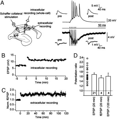
Fig. 1.
EPSPs in CA1 hippocampal neurons robustly potentiate after a single-burst stimulation of the Schaffer collateral pathway. (A) (Left) Recording configuration with whole-cell somatic patch-clamp recording, simultaneous extracellular field-potential recording, and stimulation electrode in stratum radiatum (SR, close to CA3/CA1 border). (Right) Intracellular (upper trace, resting membrane potential: −66 mV) and simultaneous extracellular (lower trace) recording of a single-burst response to a SR stimulus consisting of five pulses at 100 Hz (middle trace). (Insets) Representative EPSPs (upper) and field EPSPs (fEPSPs, lower) before (pre) and 20 min after (post) a single-burst SR stimulus. (B) Mean EPSP amplitude recorded in whole-cell patch-clamp configuration plotted vs. time. Time 0 indicates delivery of the stimulus. (C) Normalized fEPSP amplitude recorded before and after stimulation. All fEPSPs were recorded for at least 120 min (n = 4). In two of these experiments, LTP was recorded for up to 210 min (data not shown). (D) Potentiation ratio for whole-cell recordings (average EPSP 15–20 min after stimulation divided by the average of the 5-min baseline) and field potential recordings (average fEPSP 20–30 min and average fEPSP 110–120 min after stimulation divided by the average of the 10-min baseline). The potentiation ratio was not different between intracellular and extracellular recordings (ANOVA, P > 0.4). The number of experiments in each condition is indicated at bottom of the respective bars. Open circles represent the distribution of potentiation ratios in intracellular recordings (20 min after stimulation). Data are presented as mean ± SEM.

