
Fig. 6. hERβ is a strong inhibitor of motility and invasion.
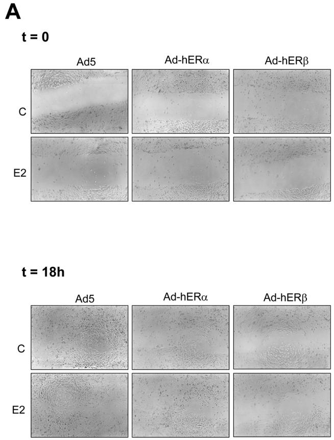
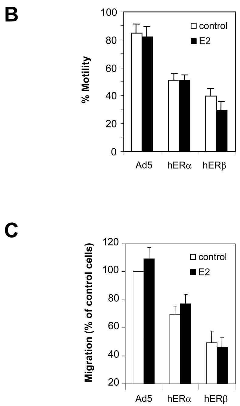
A. MDA-MB-231 cells were infected with Ad5, Ad-hERα or Ad-hERβ viruses at MOI 100. 24 h after the beginning of the infection, the cells were then treated with ethanol (control) or E2 (10−8M). After 48h of ligand treatment, cells were scratched with a blue tip and pictured (t=0). The wound was pictured again 18h after the scratch (t=18h). Pictures of a representative assay are shown here. B. Results are shown as the % of wound filling after 18h of migration and represent the mean ± SD of 3 experiments. C. MDA-MB-231 cells were infected with Ad5, Ad-hERα or Ad-hERβ (MOI 100). Cells were plated on transwell or on control plates and treated with ethanol vehicle or E2 (10−8 M) 24h after infection. Cells which have migrated to the lower side of the filter and cells present in the control plates were counted after 36h of migration. The percentage of control migrating cells was set up to 100. Results are expressed as the percentage of control migrating cells and represent the mean ± SD of four experiments.