
Fig. 1.
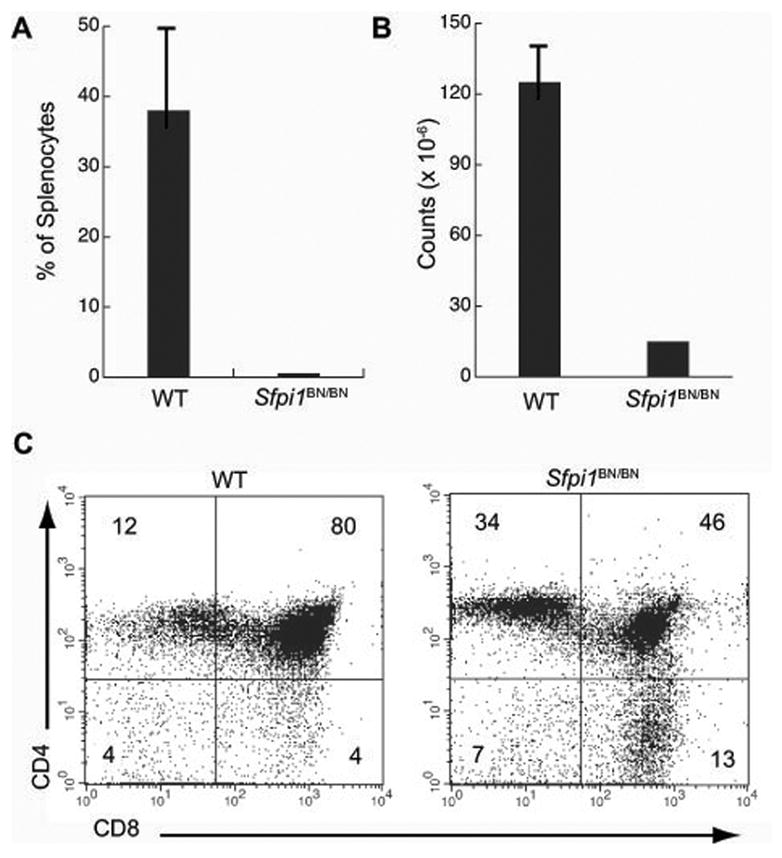
Lymphocyte development in mice homozygous for a hypomorphic allele of Sfpi1. A) Engraftment of wild type (WT) or Sfpi1BN/BN fetal liver progenitors into RAG2−/−il2rγ−/− immunodeficient mice. The percentage of B220+ CD19+ B cells in the spleen of recipient mice (n = 5) was measured by flow cytometry six weeks after i.v. injection of 2 × 10⁶ fetal liver cells. B) Total cellularity of the thymus of wild type (WT) or Sfpi1BN/BN mice. C) Flow cytometric analysis of single-cell suspensions from thymus of either wild type (WT) or Sfpi1BN/BN mice at 10 days of age. Cells were gated for proper size and granularity and incubated with an antibody to the indicated cell surface markers. Numbers represent percentages of gated cells in each quadrant.