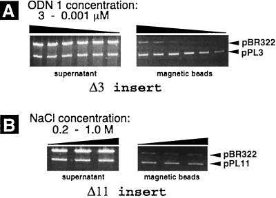
Figure 2.
Detection of the PD-loop formation by the affinity capture of linearized plasmids pPL3 and pPL11 on the streptavidin-coated magnetic beads. The gels represent DNA retained in the supernatant (Left) and DNA washed out from the magnetic beads after the affinity capture procedure (Right). The top bands in all gels correspond to the control pBR322 plasmid (without PNA binding sites), which was in 4-fold excess over pPL3 and pPL11 plasmids. (A) Effect of oligonucleotide concentration on the efficiency of capturing of the pPL3 plasmid. Complexing of ODN 1 was performed in buffer solution containing 1 M NaCl, and samples corresponding to adjacent lanes represent a 5-fold difference in the oligonucleotide concentration. (B) Effect of salt concentration on the capturing efficiency of the pPL11 plasmid. ODN 1 was at 3 μM in buffer solution containing 0.2, 0.5, and 1 M NaCl.

