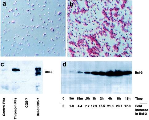
Figure 1.
Activated platelets express Bcl-3. (a and b) Immunocytochemical detection of Bcl-3. Bcl-3 was examined in resting (a) or thrombin-stimulated (b) (0.1 unit/ml for 2 hr) platelets as described in Materials and Methods. Red staining in b indicates the presence of Bcl-3 and is prominent in platelet aggregates. Rabbit IgG, or deletion of the primary antibody, did not result in red staining, indicating that the immunolocalization observed was specific for Bcl-3. (c) Detection of Bcl-3 by Western blot analysis in stimulated platelets and transfected cells. Cellular lysates from control or activated platelets (plts) stimulated with 0.1 unit/ml of thrombin for 2 hr, or from COS-7 cells transfected with vector or Bcl-3 cDNA, were obtained, and Bcl-3 expression (56-kDa protein) was determined by Western blot analysis. In transfected COS-7 cells (lane 4), the slower-migrating bands are phosphorylated forms of Bcl-3 protein that are concentrated in the cytoplasm whereas the faster-migrating band is not phosphorylated and concentrates in the nucleus (not shown). (d) Bcl-3 protein increases over time in platelets activated with thrombin. Platelets were stimulated with thrombin (0.1 unit/ml), and Bcl-3 expression was examined by Western blot analysis over 18 hr. Numbers at the bottom indicate the increase in Bcl-3 protein expression over baseline as measured by densitometry (m, minutes; h, hours). This figure is representative of three experiments.

