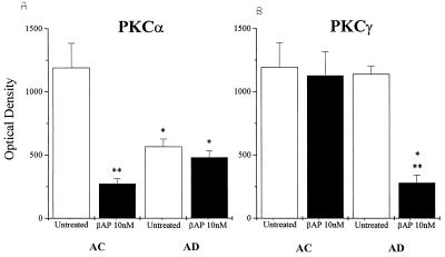
Figure 3.
Bar graphs shows that PKCα (A) and PKCγ (B) are decreased in fibroblasts from AC fibroblasts and AD fibroblasts, respectively, after treatment with 10 nM βAP(1–40) for 48 h (optical density is an arbitrary unit from densitometric analyses of the immunoreactive bands; values are the mean ± SEM of 10 or more experiments per group; ∗∗, P < 0.001, significance versus the untreated group; ∗, P < 0.001, significance versus the AC group).

