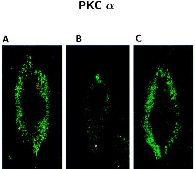
Figure 8.
Confocal microscopy imaging of the effects of βAP(1–40) on PKCα immunofluorescence in AC fibroblasts. (A) PKCα immunofluorescence was localized in the perinuclear area of nontreated AC fibroblasts. (B) PKCα signal is abolished in AC cell after treatment with 10 nM βAP(1–40) for 48 h. (C) PKCα immunofluorescence was almost completely restored after PKC activation with 100 nM PMA for 30 min in βAP-treated AC.

