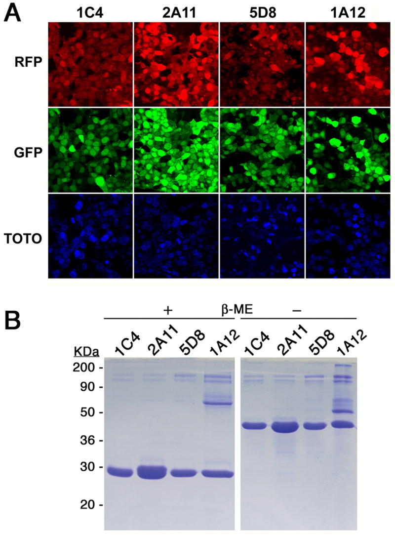
Fig. 2.

Fluorescence levels, yields and folding of expressed Fabs from production cell lines. (A) Cells secreting a specific Fab fragment, 1C4, 2A11, 5D8 or 1A12, as indicated on the top of each corresponding column, were visualized by confocal microscopy for fluorescence from RFP (top row), GFP (middle row) and TOTO-3 (bottom row). (B) The different Fabs were run, after purification, on an SDS-PAGE denaturing gel after treatment with loading dye with (+) and without (−) β-mercaptoethanol. The identity of each Fab is shown above the corresponding lane.
