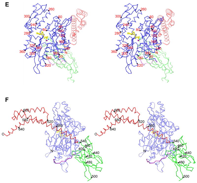
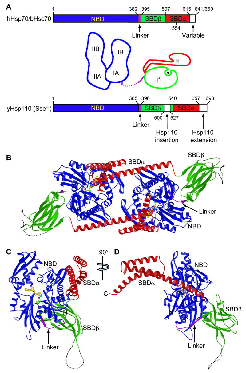
Figure 1. Overall structure of Sse1.

(A) Schematics of Hsp70 and Hsp110 sequences and prototypic Hsp70 domain structures. Coloring is NBD (blue), interdomain linker (purple), SBDβ (green), SBDα (red). (B) Ribbon diagram of the dimer. Protomer A is left and B is right. Missing loops are dotted. Protein coloring is as in A. ATP molecules have sticks for bonds (yellow) and balls for associated metal ions: Mg2+ (purple) and K+ (cyan). (C) Ribbon diagram of protomer B in the canonical, front-face NBD view. (D) Orthogonal view of the protomer drawn as in C, but from the right side. (E) Stereo drawing of a Cα trace of protomer B. NBD is in stick representation (full intensity) and SBD is in a continuous fine ribbon (subdued). View and coloring are as in C. Every 20th NBD Cα is marked with a black sphere and labeled. (F) Stereo drawing as in E, but viewed and colored as in D except that now SBD is at full intensity and NBD is subdued.

