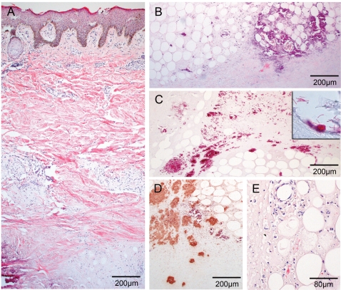
Figure 2. One patient exhibits strong histopathological signs for a progressive Buruli ulcer and large mycobacterial clumps.
Histological sections of the non-responding patient stained with HE (A, B, D) and ZN (C) or polyclonal anti-leprae antibody (pAbLep; E). Magnification ×40 (A, B, C, E) and ×100 (D). (A) Tissue shows typical signs of advanced Buruli ulcer as deep dermal necrosis, calcification or epidermal hyperplasia. Leucocytes are found in rare cases around vessels and hardly ever between fat ghosts. (B) Adipose tissue and its surroundings are highly necrotic and happen to accumulate fat ghosts and calcification. (C) The centre of the necrotic lesion harbours tremendous clumps of rod-shaped mycobacteria. (D) Staining with pAbLep demonstrates the focal clusters of live bacteria in the necrotic core. (E) If cellular infiltration occurs in small amounts within some parts of the intradermal adipose tissue cells display apoptotic features.

