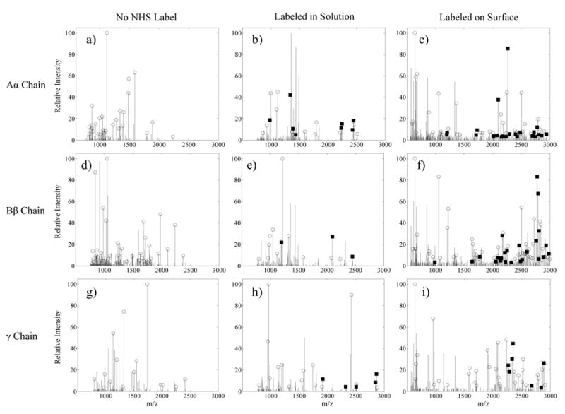
Fig. 6.

Centroided MALDI spectra corresponding to bFg trypsin digest fragment masses from the 0.5 mg/mL adsorption experiments. Open circles indicate the identification of masses that correspond to unlabeled bFg trypsin fragments. Black squares mark peaks that match an expected peptide after the Sulfo-NHS-LC-Biotin label was added to the mass. Spectra are shown for: a) Unlabeled bFg Aα chain, b) Aα chain labeled with biotin, c) Aα chain labeled with biotin after adsorption onto PET, d) Unlabeled Bβ chain, e) Bβ chain labeled with biotin, f) Bβ chain labeled with biotin after adsorption onto PET, g) Unlabeled γ chain, h) γ chain labeled with biotin, i) γ chain labeled with biotin after adsorption onto PET.
