
Figure 1.
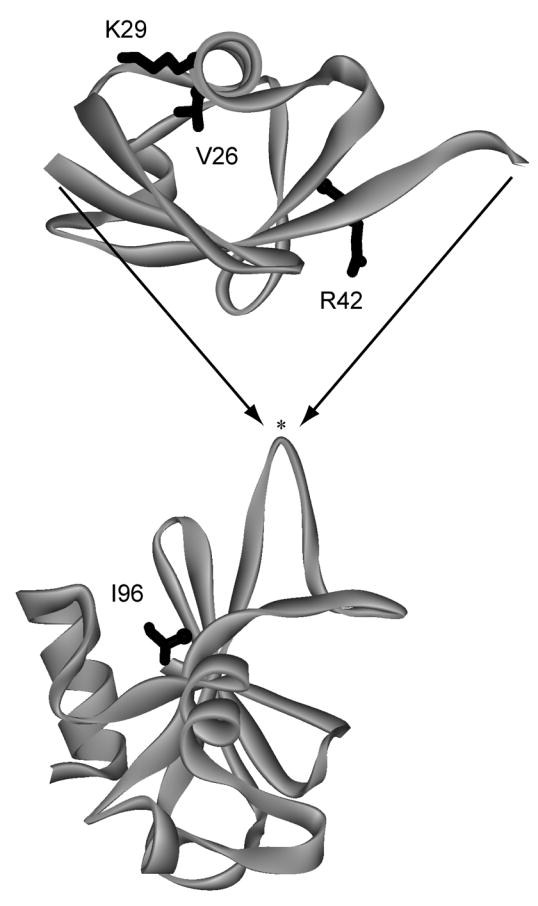
Design of the BU fusion protein. Ub (top) was inserted into a surface loop of Bn (bottom) between residues 66 and 67 (indicated by asterisk). Locations of mutated residues are shown in black. BU variants were cloned, expressed, and purified as described previously 2.